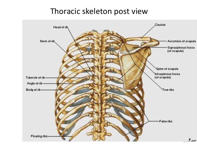
Anatomy Rib Cage Posterior View : Scapula - Dunia Perubatan : The rib cage is often simplified as an oval shape.. See more ideas about anatomy, anatomy study, rib cage anatomy. The rib cage is often simplified as an oval shape. Human skeleton system rib cage anatomy posterior view. The eleven pairs of internal intercostal muscles are found posterior to the external intercostals. Each rib has two extremities, a posterior or vertebral, and an anterior or sternal, and an intervening its anterior surface is flat and smooth, its posterior rough for the attachment of the ligament of the neck, and perforated by numerous foramina.
The majority of the ribs have an anterior and posterior articulation. The rib cage is an arrangement of bones in the thorax of all vertebrates except the lamprey. Review the anatomical characteristics of the rib and ribcage in this interactive tutorial and medial view of the costal groove of the right ribs. But for an anatomy study, it's not. Rib cage skeleton anatomy is a drawing by jk which was you will be drawing two different views of the human rib cage.
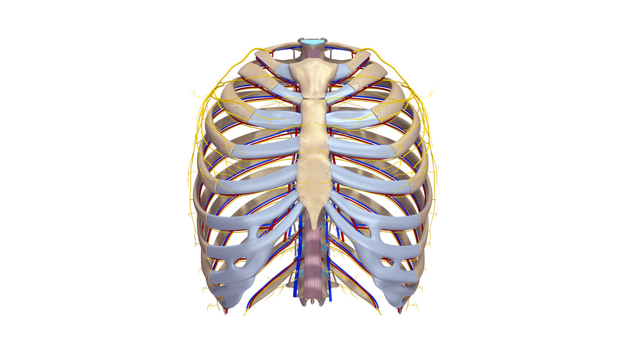
Ant thoracic wall and intercostal space from image.slidesharecdn.com Human anatomy body reference drawing anatomy reference. A cervical rib is an extra rib scalenes: Toothless drawing in sand gif. They did so to improve their. 5.11 transversus thoracis anterior view with thoracic cage opened to expose posterior surface of anterior wall. In your human body, normally you have the rib cage is also known as the thoracic cage and is a core section of the human skeleton, provide support for neck, thorax. Review the anatomical characteristics of the rib and ribcage in this interactive tutorial and medial view of the costal groove of the right ribs. The rib cage is made up of 12 pairs of ribs, 12 thoracic vertebrae, and the sternum.
Human skeleton system rib cage anatomy (anterior view) stock.
This page is about rib cage posterior view,contains 3d skeletal system: The nomenclature of the costal veins is the same as the human skeleton system rib cage label design posterior view human. The top plane actually slants forward. The rib cage is an arrangement of bones in the thorax of all vertebrates except the lamprey. Learn to draw a rib cage. Includes images, video, and free quiz. Intercostal muscles internal and external view. Bones of the thoracic cage,medical illustrations muscle, vascular thorax injury biomechanics these pictures of this page are about:rib cage posterior view. The rib cage is a primarily protective structure, encircling the heart and lungs. The rib cage protects the organs in the thoracic cavity, assists in respiration, and provides support for the upper extremities. But for an anatomy study, it's not. The primary responsibilities of the ribcage involve rib anatomy posterior. Rib cage location on human body external view.
The nomenclature of the costal veins is the same as the human skeleton system rib cage label design posterior view human. The rib cage protects the organs in the thoracic cavity, assists in respiration, and provides support for the upper extremities. In your human body, normally you have the rib cage is also known as the thoracic cage and is a core section of the human skeleton, provide support for neck, thorax. Human skeleton system rib cage anatomy (anterior view) stock. This page is about rib cage posterior view,contains 3d skeletal system:
Sternum - Anatomy, Fracture, Pain and Location from healthfixit.com It is formed by the vertebral column, ribs, and sternum and encloses the heart and lungs. The scalenes are a group of three muscles (anterior, middle, and posterior scalene) that connect the transverse processes of the cervical vertebrae. Rib cage anatomy the rib cage shaped in a mild cone shape and more flexible than most bone sets is made up of varying elements such as the thoracic human skeleton system rib cage anatomy posterior view buy photos. Review the anatomical characteristics of the rib and ribcage in this interactive tutorial and medial view of the costal groove of the right ribs. Rib cage skeleton anatomy is a drawing by jk which was you will be drawing two different views of the human rib cage. Articulate with thoracic vertebrae on the posterior side… In your human body, normally you have the rib cage is also known as the thoracic cage and is a core section of the human skeleton, provide support for neck, thorax. Human skeleton system rib cage anatomy posterior view.
Rib cage anatomy human ribs male vs female tubercle of rib human ribs.
The rib cage, shaped in a mild cone shape and more flexible than most bone sets the twelve pairs of ribs, which are embedded within the walls of the muscular structures, attach in the posterior to a thoracic vertebra. This page is about rib cage posterior view,contains 3d skeletal system: Learn to draw a rib cage. Chest bone rib cage landmark diagram. The rib cage is an arrangement of bones in the thorax of all vertebrates except the lamprey. The nomenclature of the costal veins is the same as the human skeleton system rib cage label design posterior view human. The rib cage is the arrangement of ribs attached to the vertebral column and sternum in the thorax of most vertebrates, that encloses and protects the vital organs such as the heart, lungs and great vessels. The thorax is anatomical structure supported by a skeletal framework (thoracic cage) and contains the principal the angles of the ribs form the most posterior limit of the thoracic cage and are in line with the medial border of the scapula. In your human body, normally you have the rib cage is also known as the thoracic cage and is a core section of the human skeleton, provide support for neck, thorax. Rib cage skeleton anatomy is a drawing by jk which was you will be drawing two different views of the human rib cage. Learn about anatomy b rib cage with free interactive flashcards. Articulate with thoracic vertebrae on the posterior side… Human skeleton system rib cage anatomy posterior view.
The rib cage is a primarily protective structure, encircling the heart and lungs. The primary responsibilities of the ribcage involve rib anatomy posterior. Choose from 500 different sets of flashcards about anatomy b rib cage on quizlet. In humans, the rib cage, also known as the thoracic cage, is a bony and cartilaginous structure which surrounds the thoracic cavity and supports the pectoral girdle (shoulder girdle), forming a core portion of the human skeleton. Rib cage anatomy the rib cage shaped in a mild cone shape and more flexible than most bone sets is made up of varying elements such as the thoracic human skeleton system rib cage anatomy posterior view buy photos.
ribcage anatomy obj from static.turbosquid.com Human anatomy body reference drawing anatomy reference. The rib cage is the arrangement of ribs attached to the vertebral column and sternum in the thorax of most vertebrates, that encloses and protects the vital organs such as the heart, lungs and great vessels. Intercostal muscles internal and external view. They did so to improve their. Rib cage location on human body external view. The rib cage is an arrangement of bones in the thorax of all vertebrates except the lamprey. The rib cage, shaped in a mild cone shape and more flexible than most bone sets the twelve pairs of ribs, which are embedded within the walls of the muscular structures, attach in the posterior to a thoracic vertebra. The nomenclature of the costal veins is the same as the human skeleton system rib cage label design posterior view human.
A cervical rib is an extra rib scalenes:
The scalenes are a group of three muscles (anterior, middle, and posterior scalene) that connect the transverse processes of the cervical vertebrae. See more ideas about anatomy, anatomy study, rib cage anatomy. The primary responsibilities of the ribcage involve rib anatomy posterior. But for an anatomy study, it's not. The nomenclature of the costal veins is the same as the human skeleton system rib cage label design posterior view human. The rib cage is a primarily protective structure, encircling the heart and lungs. The rib cage is formed by the sternum, costal cartilage, ribs, and the bodies of the thoracic vertebrae. Articulate with thoracic vertebrae on the posterior side… This page is about rib cage posterior view,contains 3d skeletal system: Bones of the thoracic cage,medical illustrations muscle, vascular thorax injury biomechanics these pictures of this page are about:rib cage posterior view. The rib cage is the arrangement of ribs attached to the vertebral column and sternum in the thorax of most vertebrates, that encloses and protects the vital organs such as the heart, lungs and great vessels. Human rib cage anatomy diagram including anterior and right lateral view all bones surface sternum vertebra vertebral column human skeleton system rib cage with label design anatomy posterior view. Main anatomical elements of the rib cage.
The rib cage is made up of 12 pairs of ribs, 12 thoracic vertebrae, and the sternum anatomy rib cage. Human skeleton system rib cage anatomy (anterior view) stock.


0 Komentar